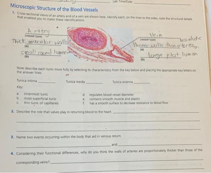
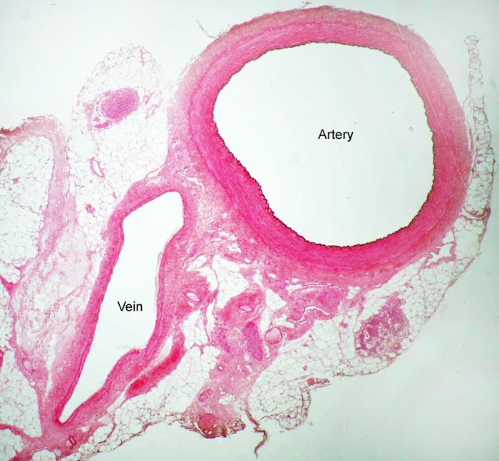
Cross sectional views of an artery and of a vein – Embark on a captivating exploration of cross-sectional views of arteries and veins, revealing the intricate histological architecture and functional significance that shape the circulatory system. This comprehensive overview unravels the structural distinctions, histological layers, and functional implications of these vital vessels, providing a deeper understanding of their roles in maintaining cardiovascular health.
Delving into the microscopic realm, we dissect the histological features of arteries and veins, examining the tunica intima, tunica media, and tunica adventitia. These layers orchestrate the flow of blood, ensuring efficient oxygen and nutrient delivery throughout the body.
Cross-Sectional Views of an Artery and a Vein: General Overview
Cross-sectional views of arteries and veins provide valuable insights into their structural and functional differences. Arteries carry oxygenated blood away from the heart, while veins carry deoxygenated blood back to the heart. These vessels exhibit distinct histological features and wall compositions that reflect their specific roles in the circulatory system.
Differences in Wall Thickness and Composition
Arteries have thicker walls compared to veins due to the presence of a well-developed tunica media. The tunica media contains smooth muscle cells that allow for vasoconstriction and vasodilation, regulating blood flow to tissues. In contrast, veins have thinner walls with a less prominent tunica media.
This difference in wall thickness contributes to the functional distinctions between arteries and veins.
Functional Significance of Differences
The thicker walls of arteries enable them to withstand higher blood pressure generated by the heart’s pumping action. This ensures adequate perfusion of oxygenated blood to tissues. Conversely, the thinner walls of veins allow for lower blood pressure and facilitate the return of blood to the heart.
Additionally, valves present in veins prevent backflow of blood and assist in maintaining venous blood flow against gravity.
Histological Features of Arteries

Tunica Intima
The tunica intima is the innermost layer of an artery. It consists of a single layer of endothelial cells resting on a basement membrane. The endothelial cells play a crucial role in regulating vascular tone, inflammation, and thrombosis.
Tunica Media
The tunica media is the middle layer of an artery and is composed primarily of smooth muscle cells arranged in concentric layers. These smooth muscle cells control the diameter of the artery through vasoconstriction and vasodilation, adjusting blood flow to meet tissue demands.
Tunica Adventitia
The tunica adventitia is the outermost layer of an artery and consists of connective tissue, fibroblasts, and collagen fibers. It provides structural support and protection for the artery.
Histological Features of Veins

Tunica Intima
Similar to arteries, the tunica intima of a vein consists of a single layer of endothelial cells supported by a basement membrane. However, veins may have additional layers of endothelial cells, depending on their size and location.
Tunica Media
The tunica media of a vein is thinner than that of an artery and contains fewer layers of smooth muscle cells. This reduced smooth muscle content contributes to the lower blood pressure in veins compared to arteries.
Tunica Adventitia
The tunica adventitia of a vein is composed of connective tissue and collagen fibers, similar to arteries. However, veins often have a thicker adventitia, providing additional support and protection.
Comparison of Arteries and Veins
Arteries have thicker walls with a prominent tunica media, enabling them to withstand higher blood pressure. Veins have thinner walls with a less developed tunica media, facilitating lower blood pressure and the return of blood to the heart. Additionally, veins possess valves to prevent backflow of blood.
Functional Differences between Arteries and Veins: Cross Sectional Views Of An Artery And Of A Vein

Blood Flow and Pressure
Arteries carry oxygenated blood away from the heart at high pressure, ensuring adequate perfusion of tissues. Veins carry deoxygenated blood back to the heart at lower pressure, facilitated by valves that prevent backflow.
Structural Contributions to Functional Differences, Cross sectional views of an artery and of a vein
The thicker walls and well-developed tunica media of arteries enable them to withstand high blood pressure. The thinner walls and less prominent tunica media of veins allow for lower blood pressure and facilitate venous return.
Impact on Circulatory System
The functional differences between arteries and veins are crucial for maintaining proper blood flow and pressure throughout the circulatory system. Arteries ensure the delivery of oxygenated blood to tissues, while veins facilitate the return of deoxygenated blood to the heart for reoxygenation.
FAQ Insights
What are the key structural differences between arteries and veins?
Arteries possess thicker walls composed of more layers of smooth muscle, while veins have thinner walls with less smooth muscle.
How do the functional differences between arteries and veins impact the circulatory system?
Arteries carry oxygenated blood away from the heart, while veins return deoxygenated blood to the heart. This pressure gradient ensures efficient circulation.
What are the clinical applications of cross-sectional views of arteries and veins?
These views are used in angiography and ultrasound to diagnose and monitor cardiovascular diseases such as atherosclerosis and aneurysms.